DIETARY SUPPLEMENTS

Découvrez 08 - Slim
Your ally for a slimmer figure and healthy weight

Journal
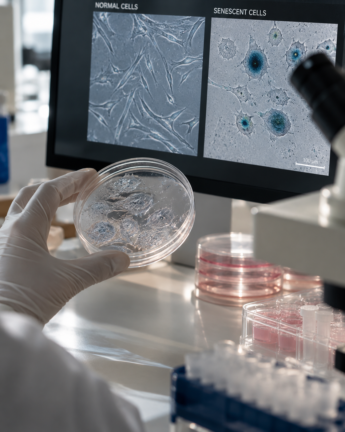
What if some aging cells didn’t die—but instead remained in our tissues, gradually changing the environment around them?
Over the past few years, one term has moved to the forefront of longevity research:
cellular senescence.
Along with it, a new vocabulary has made its way from research laboratories into the worlds of longevity, dietary supplements, skincare and healthy aging:
senescent cells.
Senolytics.
Senomorphics.
Fisetin.
Quercetin.
Rapamycin.
Dasatinib + quercetin.
And with them, an extraordinary promise:
eliminating the cells that make us age.
It is a fascinating idea.
It is also far more complex than that promise suggests.
A senescent cell is not simply an:
“old cell.”
It is not dead.
It is not necessarily harmful in every context.
And most importantly:
cellular senescence is not inherently bad.
Senescence is a biological mechanism that can help prevent damaged cells from continuing to proliferate and can play a role in certain physiological and tissue responses.
The problem appears to arise when some senescent cells:
persist.
They can remain metabolically active and gradually alter their environment through the signals they release.
That distinction changes everything.
Science is not simply asking:
“How can we stop our cells from becoming senescent?”
Researchers are trying to understand:
Why do some senescent cells persist as we age?
What do they secrete?
How do they affect surrounding cells?
Do they contribute to skin aging?
Can they be selectively eliminated?
And, perhaps most importantly:
If we eliminate them, do we actually rejuvenate the tissue?
In 2013, a landmark scientific paper proposed nine major biological mechanisms of aging—the now-famous:
Hallmarks of Aging.
Ten years later, in 2023, Carlos López-Otín, Maria Blasco, Linda Partridge, Manuel Serrano and Guido Kroemer published a major update to this framework in Cell [1].
The expanded framework now includes twelve hallmarks:
genomic instability;
telomere attrition;
epigenetic alterations;
loss of proteostasis;
disabled macroautophagy;
deregulated nutrient sensing;
mitochondrial dysfunction;
cellular senescence;
stem cell exhaustion;
altered intercellular communication;
chronic inflammation;
dysbiosis [1].
What makes this framework particularly important is that these mechanisms do not operate independently.
They interact.
Mitochondrial dysfunction can influence oxidative stress.
DNA damage can promote cellular senescence.
Senescent cells can alter intercellular communication.
Their secretory activity can contribute to an inflammatory environment.
Inflammation, in turn, can affect cellular function.
Biological aging therefore looks less like:
a clock
and more like:
a network.
And cellular senescence sits at the intersection of several of these mechanisms [1].
A senescent cell is generally defined as a cell that has entered a state characterized, among other features, by:
a durable cell-cycle arrest.
It no longer proliferates normally.
But unlike a dead cell:
it remains biologically active.
That distinction is fundamental.
A cell can therefore:
stop dividing
without:
disappearing.
It can continue to consume energy.
Continue to communicate.
Continue to produce proteins.
Continue to influence its environment.
And depending on the cell type and biological context, it can develop a distinctive secretory profile known as the:
SASP — Senescence-Associated Secretory Phenotype [2].
This is one of the central concepts in the biology of cellular senescence.
Because continuing to divide is not always the safest option.
Cells can be exposed to many different forms of stress:
DNA damage;
telomere dysfunction;
oxidative stress;
oncogene activation;
mitochondrial stress;
radiation;
ultraviolet exposure;
certain cytotoxic treatments;
and other disruptions severe enough to trigger a sustained cellular response [1–3].
Imagine a cell whose DNA has accumulated significant damage.
Two outcomes would be particularly problematic:
continuing to proliferate despite that damage
or:
passing those abnormalities on to daughter cells.
The body therefore has several mechanisms that can restrict the proliferation of a compromised cell.
Among them:
apoptosis
and:
cellular senescence.
Apoptosis is a form of programmed cell death.
The cell is eliminated through a tightly controlled biological process.
Senescence works differently.
The cell can remain within the tissue while entering a durable state of proliferative arrest.
In simplified terms:
APOPTOSIS
→ the cell is eliminated.
CELLULAR SENESCENCE
→ the cell remains, but no longer proliferates normally.
This difference explains much of what makes senescent cells so biologically interesting.
Because a cell that remains within a tissue:
continues to exist within that tissue.
And therefore may continue:
interacting with it.
The phrase:
“zombie cells”
has become extremely popular in longevity marketing.
It is memorable.
Biologically, however, it is imprecise.
A senescent cell is not:
half-dead.
Quite the opposite.
It remains:
metabolically active.
It can exert significant biological effects.
It can alter its microenvironment.
It can interact with the immune system.
And its phenotype can vary considerably depending on:
the cell type,
the tissue,
the trigger that induced senescence,
the length of time the cell has been senescent,
and the surrounding microenvironment.
“Zombie cell” therefore dramatically oversimplifies a highly complex biological state.
The scientifically accurate term is:
senescent cell.
Here lies the first paradox.
We are going to spend much of this article examining how the accumulation of senescent cells may contribute to aging.
Yet the mechanism itself can also serve protective functions.
Cell-cycle arrest can act as a barrier against the proliferation of certain damaged or oncogenically activated cells.
Under some circumstances, a potentially dangerous cell that permanently stops dividing is preferable to a potentially dangerous cell that continues to replicate.
Cellular senescence therefore contributes to mechanisms of:
tumor suppression [1,3].
Senescence can also occur transiently as part of certain physiological and tissue responses.
Which is why the scientific objective cannot simply be:
ELIMINATE SENESCENCE.
Doing so would ignore an essential part of its biology.
Potentially when its timing and context change.
At a simplified level, we can distinguish between:
transient senescence
and:
persistent senescence.
In some situations, a cell becomes senescent, performs a biological function and may subsequently be cleared, in part through immune surveillance.
But with aging, some senescent cells may:
accumulate.
The immune system’s ability to recognize and clear them may also change with age.
As a result, persistent senescent cells can continue sending signals into their surrounding environment [2,3].
This brings us to:
the SASP.
SASP stands for:
Senescence-Associated Secretory Phenotype.
The term describes a collection of molecules secreted by certain senescent cells.
Depending on cell type and context, the SASP can include:
cytokines;
chemokines;
growth factors;
proteases;
extracellular matrix-remodeling factors [2,3].
Molecules frequently investigated in different SASP models include:
IL-6;
IL-8;
certain MMPs — matrix metalloproteinases;
and many other mediators.
But one distinction is essential:
there is no single universal SASP.
The SASP of a senescent fibroblast is not necessarily identical to that of a senescent melanocyte.
Senescence induced by radiation can differ from senescence induced by other forms of stress.
And the secretory phenotype itself can evolve over time.
Cellular senescence is therefore:
a heterogeneous biological state.
Not a universal:
ON/OFF switch.
Yes.
And this may be one of the most fascinating aspects of cellular senescence.
Molecules released by certain senescent cells can alter the behavior of neighboring cells.
This is known as:
paracrine signaling
and, in certain experimental contexts:
paracrine senescence.
In other words, a cell that has experienced a particular stress can potentially alter its environment enough to affect cells that were not initially exposed to the same stress.
This phenomenon has been studied particularly closely in human skin.
A 2019 study published in The EMBO Journal examined the role of senescent melanocytes in human skin aging [4].
Among the samples studied, the researchers found that melanocytes represented the epidermal population expressing the senescence-associated marker:
p16INK4A
during skin aging.
These melanocytes also displayed other senescence-associated features, including:
reduced HMGB1
and:
dysfunctional telomeres.
And here is a particularly important finding:
those telomeres were dysfunctional without detectable telomere shortening [4].
That distinction matters.
Because it reminds us that:
telomere dysfunction does not necessarily mean short telomeres.
In the same study, researchers showed experimentally that the SASP produced by senescent melanocytes could induce telomere dysfunction in neighboring cells and limit their proliferation [4].
The mechanism involved, among other factors:
IP-10/CXCL10;
its receptor:
CXCR3;
and increased:
mitochondrial ROS.
In three-dimensional human epidermal equivalents, senescent melanocytes also impaired basal keratinocyte proliferation and contributed to an epidermal atrophy phenotype [4].
In other words:
a senescent cell can alter the behavior of the cells around it.
This profoundly changes the way we can think about skin aging.
Aging skin may not simply be a collection of individual cells growing older independently.
It may also be:
an ecosystem of cells influencing one another.
We need to be very careful with that wording.
Saying:
“aging is contagious between cells”
would be dramatic.
But scientifically, it would be excessive.
What some experimental models show is that signals released by senescent cells can induce alterations in neighboring cells.
These can include:
oxidative stress;
telomere dysfunction;
changes in proliferation;
and, in certain models, features of secondary senescence [4].
The more accurate term is therefore:
paracrine effects of cellular senescence.
Not:
“contagious aging.”
Two names appear again and again in cellular senescence research:
p16INK4A
and:
p21CIP1.
They are proteins encoded respectively by:
CDKN2A
and:
CDKN1A.
These proteins are involved in regulating the cell cycle.
In simplified terms, two major pathways frequently involved in establishing or maintaining senescence-associated cell-cycle arrest are:
p53 → p21
and:
p16 → RB.
This is why p16 and p21 expression is commonly examined in studies of cellular senescence.
But there is a problem.
There is currently:
no single, universal and perfectly specific biomarker of cellular senescence.
A cell expressing p16 is not automatically senescent in every biological context.
The same applies to p21.
And to:
SA-β-gal — senescence-associated beta-galactosidase.
Robust identification of cellular senescence generally requires:
multiple complementary markers
interpreted within a specific biological context [2,3].
Researchers may assess:
p16;
p21;
SA-β-gal;
persistent DNA damage;
γH2AX;
telomere-associated damage;
Lamin B1;
cell-cycle arrest;
cell morphology;
SASP components.
This distinction becomes particularly important when we examine senolytic studies.
Because:
lower p16 does not automatically mean “rejuvenation.”
Modern technologies such as:
single-cell RNA sequencing
and:
spatial transcriptomics
now allow scientists to investigate aging human skin at unprecedented resolution.
A recent study mapped senescence-associated signatures across the human epidermis and dermis [5].
Researchers developed a skin-specific transcriptomic signature associated with cellular senescence and identified differences between:
chronological aging
and:
photoaging.
One particularly interesting finding was that:
photoaged skin showed a greater burden of senescence-associated cellular signatures than chronologically aged skin in the analyses performed [5].
UV exposure is therefore associated not only with:
dark spots,
wrinkles,
and collagen breakdown.
It is also associated with changes in:
cellular state itself.
Ultraviolet radiation can promote:
oxidative stress;
DNA damage;
mitochondrial dysfunction;
telomere alterations;
inflammatory responses.
Depending on the intensity and biological context, a cell may:
repair the damage,
die,
or enter a senescent state.
This connects several dimensions of skin aging that are often discussed separately.
Photoaging is not simply:
a collagen problem.
It also involves:
the genome;
mitochondria;
telomeres;
inflammation;
cell-to-cell communication;
cellular senescence.
Now we return to one of the most important cells in skin biology:
the dermal fibroblast.
Fibroblasts help synthesize and remodel the extracellular matrix of the dermis.
That matrix contains:
collagens;
elastic fibers;
glycosaminoglycans;
and numerous structural and regulatory proteins.
With age, some fibroblasts develop senescence-associated characteristics.
And this transformation can have two major consequences.
First:
their own function changes.
Second:
the signals they send into the tissue change [3,5].
The exact phenotype varies by model and biological context.
But senescent fibroblasts can show changes involving:
extracellular matrix production;
inflammatory pathways;
oxidative stress;
mitochondrial function;
cell-to-cell communication;
extracellular matrix-remodeling enzymes [3].
MMPs are particularly important.
Matrix metalloproteinases — MMPs
are enzymes involved in extracellular matrix remodeling.
They are essential to normal physiology.
But excessive or dysregulated activity of certain MMPs can promote the breakdown of extracellular matrix components.
In several models of skin cell senescence, SASP profiles include increased levels of certain MMPs [3].
This creates an important biological connection:
CELLULAR SENESCENCE
↓
SASP
↓
↑ certain inflammatory mediators + ↑ certain MMPs
↓
extracellular matrix remodeling
↓
changes in the fibroblast and collagen environment
But an essential caveat remains:
collagen aging cannot be explained solely by senescent cells.
Photoaging, glycation, mechanical changes in the extracellular matrix, oxidative stress and numerous other mechanisms also contribute.
Cellular senescence is:
one piece of the puzzle.
Not:
the entire puzzle.
Recent mapping of human skin suggests that certain dermal fibroblast populations displaying senescence-associated signatures are also associated with reduced expression of genes involved in collagen and elastic fiber synthesis [5].
This is particularly interesting.
Because it suggests a potential two-sided imbalance:
less support for matrix production
combined with:
more signals capable of promoting matrix remodeling or degradation.
In simplified terms:
↓ BUILD
↑ BREAKDOWN
But again, the biology is more complex than a single equation.
Dermal fibroblasts themselves comprise multiple subpopulations.
And skin aging progressively alters their composition, microenvironment and function.
Mitochondria are often described as:
“the powerhouses of the cell.”
That is true.
But incomplete.
Mitochondria are also involved in:
cell signaling;
metabolism;
redox regulation;
apoptosis;
immunity;
and multiple stress responses.
Mitochondrial dysfunction is itself recognized as one of the Hallmarks of Aging [1].
And it is closely interconnected with cellular senescence.
Mitochondrial dysfunction can alter:
ROS production,
metabolism,
stress signaling,
inflammatory responses.
Conversely, senescence can be accompanied by profound changes in cellular metabolism and mitochondrial function.
So this is not simply:
damaged mitochondria → senescence.
It is better understood as:
a bidirectional relationship.
Once again, marketing has dramatically simplified the biology.
Reactive Oxygen Species — ROS
are not simply toxic waste products that need to be eliminated.
At physiological levels, they participate in normal cellular signaling.
The problem arises when their production and regulation become imbalanced.
This is known as:
oxidative stress.
In cellular senescence, ROS can contribute to mechanisms involving:
DNA damage;
telomere dysfunction;
stress signaling;
mitochondrial function.
The study on senescent melanocytes provides a particularly elegant example of how paracrine signaling can increase mitochondrial ROS in neighboring cells [4].
Telomeres protect the ends of chromosomes.
As cells divide and encounter different forms of stress, telomere integrity can become compromised.
Telomere shortening is one mechanism that can contribute to cellular senescence.
But another important distinction emerges:
a telomere can become dysfunctional without necessarily becoming critically short.
Persistent damage at telomeres can produce:
Telomere-Associated DNA Damage Foci — TAF.
In the study of aged human melanocytes, dysfunctional telomeres were observed:
without detectable telomere shortening [4].
This finding challenges another oversimplified equation:
LONG TELOMERES = YOUNG
SHORT TELOMERES = OLD.
The real biology is far more nuanced.
The term:
inflammaging
describes the chronic, low-grade inflammatory state associated with aging.
In the 2023 update of the Hallmarks of Aging, chronic inflammation is itself recognized as a hallmark [1].
Cellular senescence may contribute to this environment.
Some senescent cells produce SASPs rich in pro-inflammatory mediators.
When these cells persist, their signaling can contribute to maintaining an inflammatory microenvironment locally and, depending on the context, potentially more broadly.
But it would be an oversimplification to say:
“senescent cells cause inflammaging.”
Inflammaging is multifactorial.
It involves changes in:
immune function,
metabolism,
mitochondria,
the microbiome,
tissue homeostasis.
Cellular senescence is one mechanism that may contribute to it.
In certain contexts, conceptually, yes.
We can imagine:
CELLULAR STRESS
↓
CELLULAR SENESCENCE
↓
SASP
↓
INFLAMMATORY SIGNALING
↓
STRESS IN NEIGHBORING CELLS
↓
TISSUE DYSFUNCTION
But this is not necessarily a simple linear cascade.
It is better understood as a network of feedback loops.
And this is precisely what the modern Hallmarks of Aging framework suggests:
the mechanisms of aging interact with and reinforce one another [1].
Two broad mechanisms may coexist.
1. We may generate more of them.
Over time, cells experience increasing exposure to:
damage,
stress,
mitochondrial dysfunction,
telomere alterations,
environmental insults.
2. We may become less efficient at clearing them.
The immune system contributes to the surveillance and removal of certain senescent cells.
But immune function itself changes with age.
The potential result is straightforward:
increased production + reduced clearance = accumulation.
Yet accumulation remains highly dependent on:
the tissue,
the cell type,
age,
physiological state,
disease,
environment.
No.
And this may be one of the most important facts shaping the future of senolytic therapies.
Senescent cells can arise:
in different tissues,
from different cell types,
in response to different stressors,
with different SASPs,
and with different survival dependencies.
A:
senescent fibroblast
is not necessarily biologically equivalent to:
a senescent melanocyte
or:
a senescent adipocyte.
This heterogeneity creates a major challenge.
Because a treatment capable of killing one senescent cell population:
may not kill another.
This is one reason why the idea of:
“THE senolytic”
probably makes little biological sense.
We have reached the central paradox.
Cellular senescence can:
protect us.
It can stop a damaged cell from proliferating.
It can participate transiently in certain biological processes.
But when a senescent cell:
persists;
resists clearance;
accumulates;
continues producing a harmful SASP;
the same biological strategy can become problematic [2,3].
The question may therefore not be:
“How do we stop cellular senescence?”
But rather:
“How do we eliminate the right senescent cells at the right time without disrupting the useful functions of senescence?”
And this is precisely where an entirely new category of compounds enters the picture:
SENOLYTICS.
The idea almost sounds too simple.
A senescent cell accumulates.
It releases potentially harmful signals.
So:
eliminate it.
But one question immediately arises:
How do you kill a senescent cell without killing the healthy cells around it?
To answer that question, researchers began investigating a particularly intriguing feature of certain senescent cells.
They can become:
resistant to apoptosis.
In other words:
they no longer proliferate,
they may be damaged or profoundly altered,
yet they activate certain mechanisms that allow them to:
survive.
These mechanisms are sometimes grouped under the term:
Senescent Cell Anti-Apoptotic Pathways — SCAPs.
What if we could disrupt those survival mechanisms?
The senescent cell could become more vulnerable to apoptosis.
That is the principle behind:
SENOLYTICS.
But there is another strategy.
Instead of killing the cell:
what if we could simply make it less harmful?
That is the principle behind:
SENOMORPHICS.
In Part 2, we will put this theory up against the actual experimental and human evidence.
Dasatinib + quercetin.
Fisetin.
Rapamycin.
Navitoclax.
Why are some of these compounds considered senolytics while others are better described as senomorphics?
Is fisetin really a:
“natural senolytic”?
Does the quercetin now found in countless longevity supplements actually clear senescent cells in humans?
What happened when researchers administered senolytics to:
9 patients with diabetic kidney disease?
To people with:
idiopathic pulmonary fibrosis?
To:
60 postmenopausal women
in a randomized clinical trial?
And most importantly for skin longevity:
what happens when this strategy is tested specifically on skin and collagen?
Researchers have now conducted experiments using:
actual aged human skin.
With intriguing findings involving:
senescent cells;
SASP;
MMPs;
inflammation;
collagen density.
But with one crucial methodological distinction:
human skin does not necessarily mean a human clinical trial.
Finally, we will address the question longevity marketing often prefers to skip:
Even if senolytics work biologically, have we actually shown that they slow human aging?
A senolytic is a compound designed to preferentially induce the death of certain senescent cells while sparing non-senescent cells as much as possible [6,7].
The word combines:
senescent
lytic — destruction.
The concept is based on a fascinating observation.
Some senescent cells exist in a biological paradox.
They have experienced enough stress to stop proliferating.
They may exhibit:
DNA damage,
oxidative stress,
mitochondrial dysfunction,
pro-apoptotic signaling.
And yet:
they survive.
Why?
Because certain senescent cells activate survival networks that help them resist their own pro-apoptotic environment [6].
These mechanisms are sometimes grouped under the term:
Senescent Cell Anti-Apoptotic Pathways — SCAPs.
One senolytic strategy is therefore to interfere with these survival pathways.
In simplified terms:
SENESCENT CELL
↓
ACTIVATION OF SURVIVAL PATHWAYS
↓
RESISTANCE TO APOPTOSIS
↓
SENOLYTIC
↓
DISRUPTION OF SURVIVAL SIGNALING
↓
APOPTOSIS
But this simplified model hides a fundamental challenge.
Not all senescent cells rely on the same survival mechanisms.
A senolytic that is active against:
a senescent fibroblast
may be far less effective against:
a senescent adipocyte.
Or an immune cell.
Or even a fibroblast that became senescent in response to a different type of stress.
Senolytic activity can depend on:
the cell type,
the tissue,
the mechanism that triggered senescence,
the stage of senescence,
the dose,
and the microenvironment [6].
This is one of the most important concepts in the entire field.
The term:
“senolytic”
does not describe a universal biological eraser capable of clearing every senescent cell from the body.
It describes:
a therapeutic strategy.
There is another approach.
Instead of eliminating a senescent cell, what if we could:
change its behavior?
This is the principle behind:
SENOMORPHICS
also referred to, in some classifications, as:
senostatics.
A senolytic primarily aims to:
eliminate certain senescent cells.
A senomorphic instead aims to:
reduce potentially harmful features of the senescent phenotype, particularly components of the SASP, without necessarily killing the cell [7].
The distinction can be summarized simply:
SENOLYTIC
→ kill the cell.
SENOMORPHIC
→ quiet the cell.
These represent two fundamentally different strategies for targeting cellular senescence and age-related biology.
Senescent cells no longer proliferate normally.
And senescent cells may accumulate gradually over time.
This has led researchers to investigate so-called:
“hit-and-run” strategies.
The goal is not necessarily to expose the body continuously to a senolytic drug.
In theory, intermittent treatment could eliminate a proportion of treatment-sensitive senescent cells before they accumulate again.
This approach has been investigated in several human studies involving:
dasatinib + quercetin [8–11].
But this distinction is critical:
there is currently no validated intermittent “anti-aging senolytic protocol” for healthy people.
The clinical studies discussed below were conducted in specific medical populations and research settings.
The combination:
D + Q
stands for:
DASATINIB + QUERCETIN.
It has become one of the best-known combinations in senolytic research.
And the two compounds are very different.
Dasatinib is a tyrosine kinase inhibitor used as a prescription cancer drug.
Quercetin is a flavonoid naturally found in a variety of foods and also widely sold as a dietary supplement.
The fact that quercetin occurs naturally does not make D+Q a “natural” treatment.
More importantly:
dasatinib is a prescription drug with potentially significant side effects, contraindications and drug interactions.
D+Q is an experimental strategy being investigated in clinical research.
It is not a DIY longevity protocol.
This is where the story becomes particularly interesting.
In 2019, a small phase 1 study enrolled:
9 people with diabetic kidney disease [8].
Participants received:
dasatinib + quercetin
for three days.
Researchers collected samples of:
adipose tissue;
skin;
blood
before treatment and again eleven days after the final dose [8].
They then investigated several parameters associated with cellular senescence.
The results became one of the field’s important early demonstrations of senolytic pharmacodynamic activity in humans.
In adipose tissue, researchers observed reductions in cells expressing:
p16INK4A
and:
p21CIP1.
They also observed a reduction in cells showing:
SA-β-gal activity [8].
The raw values reported in adipose tissue biopsies decreased by approximately:
35% for p16+ cells;
17% for p21+ cells;
62% for SA-β-gal+ cells [8].
But one of the most interesting findings for skin aging involved:
the skin itself.
Eleven days after treatment ended, researchers observed an approximately:
20% reduction in p16INK4A+ cells
and:
31% reduction in p21CIP1+ cells
in the basal layer of the epidermis [8].
Several circulating SASP-associated factors also decreased, including:
IL-1α;
IL-6;
MMP-9;
MMP-12 [8].
This is an important result.
It shows that a senolytic intervention in humans can be followed by reductions in several biological parameters associated with senescent cell burden.
But the essential context is:
9 participants.
All had:
diabetic kidney disease.
The study did not demonstrate that their skin developed:
fewer wrinkles,
more collagen,
greater elasticity,
or a younger clinical appearance.
It demonstrated:
biological activity.
Not:
clinical skin rejuvenation.
Taken together, the findings are consistent with a reduction in senescent cell burden, which is how the authors interpreted the results [8].
But remember one of the central principles from Part 1:
p16 is not a universal senescent-cell counter.
Neither is:
p21.
The strength of this study comes from the convergence of multiple parameters:
p16,
p21,
SA-β-gal,
replicative potential,
circulating SASP factors.
That is much more informative than a change in a single biomarker.
But these biomarkers still cannot be interpreted as a direct measurement of:
the participant’s overall biological age.
This is where the clinical literature becomes far less dramatic than longevity marketing.
An early open-label pilot study investigated D+Q in people with:
idiopathic pulmonary fibrosis — IPF [9].
Changes in certain measures of physical function were reported.
But the study:
had no placebo group.
It was small.
And it was not designed to demonstrate that D+Q slows human aging.
A subsequent pilot trial was:
randomized;
placebo-controlled;
single-blind
and included only:
12 participants [10].
Its primary focus was:
feasibility and tolerability.
It was not statistically powered to establish clinical efficacy.
Exploratory measures of frailty, pulmonary function and physical performance did not show interpretable significant differences between groups [10].
A phase 1 study also evaluated D+Q in:
5 people with early-stage symptomatic Alzheimer’s disease [11].
The primary objectives included:
central nervous system penetration;
safety;
feasibility.
Dasatinib was detected in the cerebrospinal fluid of four of the five participants.
Quercetin was not [11].
Treatment was generally tolerated in this very small group.
But cognitive and neuroimaging endpoints did not show significant changes between baseline and the end of the study [11].
Again:
5 people.
No placebo group.
No demonstration of brain rejuvenation.
In 2024, a phase 2 study investigated intermittent D+Q treatment in:
60 postmenopausal women [12].
This time, researchers had a:
randomized trial
with a defined primary endpoint.
The study focused in part on bone metabolism.
Its primary endpoint was the change at 20 weeks in:
CTx — C-terminal telopeptide of type I collagen,
a marker of bone resorption.
And the result is essential:
the primary endpoint was not met [12].
At 20 weeks, the change in CTx did not differ significantly between the D+Q and control groups [12].
A marker of bone formation:
P1NP — procollagen type I N-terminal propeptide
increased by approximately:
16% relative to control
at 2 and 4 weeks.
But that difference was no longer present at:
20 weeks [12].
In other words:
the intervention did not produce the expected primary outcome across the overall study population.
This matters enormously.
Because science advances through:
negative results, too.
The researchers also conducted:
exploratory analyses.
Among participants in the tertile with the highest baseline levels of:
p16 mRNA in T cells,
some bone-related responses appeared more favorable [12].
At two weeks, this subgroup showed an increase in P1NP and a decrease in CTx.
An increase in radial bone mineral density was also observed at 20 weeks [12].
This is intriguing.
But:
an exploratory subgroup analysis is not definitive clinical evidence.
It generates a hypothesis.
And that hypothesis is fascinating:
Could the effectiveness of a senolytic depend on a person’s baseline senescent cell burden?
Further trials will be needed to answer that question.
No.
Current evidence does not support recommending pharmacological senolytics to healthy adults for the purpose of:
slowing aging;
extending lifespan;
rejuvenating the skin;
or:
broadly preventing age-related disease [7,12].
Human trials remain:
small,
heterogeneous,
conducted in patients with disease or highly specific populations,
and often designed primarily to investigate safety, feasibility, pharmacodynamics or a particular medical indication.
The field has therefore moved beyond:
“only tested in mice.”
But it remains far from:
“proven anti-aging treatment in humans.”
A 2024 study specifically examined the effects of D+Q on:
senescent human dermal fibroblasts
and:
aged human skin [13].
Researchers induced senescence in human dermal fibroblasts using several approaches:
extended passaging,
ionizing radiation,
doxorubicin.
D+Q selectively eliminated a proportion of senescent fibroblasts in these models [13].
Then the researchers went further.
They used:
actual skin tissue from older humans.
But what happened next is crucial.
The aged human skin samples were grafted onto:
immunodeficient mice [13].
The mice then received D+Q.
Following treatment, the human skin grafts showed:
reductions in senescence-associated parameters;
reductions in SASP components;
reductions in certain MMPs and interleukins;
increased collagen density [13].
This is a particularly interesting experimental model.
Because the tissue being studied is:
human.
But the organism receiving the treatment is:
a mouse.
The study therefore does not allow us to claim:
“D+Q increases collagen in human skin.”
What it allows us to say is:
“In a model of aged human skin grafted onto mice, D+Q was associated with the elimination of senescent fibroblasts, reduced SASP-related parameters and increased collagen density.” [13]
That distinction is critical.
Navitoclax — ABT-263
is another compound investigated for its senolytic activity.
It inhibits anti-apoptotic proteins within the:
BCL-2 family.
In another study using a model of:
aged human skin grafted onto mice,
navitoclax promoted the selective elimination of senescent dermal fibroblasts [14].
Treated grafts showed:
fewer senescent cells;
lower levels of certain SASP-associated molecules;
lower levels of certain MMPs and interleukins;
and:
greater collagen density [14].
Again:
biologically compelling.
But:
not a clinically proven anti-wrinkle treatment in humans.
Because a drug capable of disrupting anti-apoptotic mechanisms can also cause significant adverse effects.
One well-established concern with navitoclax is:
thrombocytopenia — a reduction in blood platelets.
This perfectly illustrates the challenge facing senolytic medicine.
Finding a molecule capable of:
killing a senescent cell
is not enough.
It must be sufficiently selective to avoid unacceptable toxicity in other cells and tissues [6,7].
The future of senolytic therapy will therefore likely depend heavily on:
precision targeting.
Now we come to one of the most popular compounds in the longevity space:
fisetin.
Fisetin is a:
flavonoid
naturally found in several plants and foods.
Today, it appears in numerous dietary supplements marketed around:
longevity;
healthy aging;
cellular health;
senolytic support.
It is frequently described as:
“a natural senolytic.”
But what does the science actually show?
In certain preclinical models: yes.
A study published in Biogerontology examined fisetin in:
senescent human dermal fibroblasts
and:
aged human skin grafted onto mice [15].
In cellular models, fisetin preferentially eliminated senescent fibroblasts.
Researchers observed activation of apoptotic mechanisms involving:
caspase-3;
caspase-8;
caspase-9 [15].
In aged human skin grafts, treatment was associated with:
increased collagen density
and:
reduced levels of several SASP components, including certain MMPs and interleukins [15].
This represents genuine experimental senolytic activity.
But one crucial step is still missing.
That has not been demonstrated.
The study described above did not involve giving fisetin supplements to adults and then measuring their wrinkles, skin elasticity or dermal collagen.
The human skin samples were:
grafted onto mice [15].
Fisetin was administered to the animals.
We therefore cannot transform:
“senolytic activity in fibroblasts and a human skin graft model”
into:
“taking fisetin clears senescent cells from your skin.”
That would be exactly the kind of extrapolation this article is designed to avoid.
Because the transition from:
interesting biological mechanism → commercial supplement
is often much faster than the transition from:
interesting biological mechanism → robust clinical evidence.
Fisetin continues to be investigated in clinical trials in different medical contexts.
But the existence of a trial on:
ClinicalTrials.gov
or the publication of a:
clinical trial protocol
is not evidence of efficacy.
Randomized phase 2 trials continue to be designed specifically to determine whether fisetin can produce meaningful clinical benefits in selected populations [16].
If the question is still being tested:
the answer is not already known.
Quercetin also requires considerable nuance.
It occurs naturally in many foods.
And there is an extensive scientific literature on its effects involving:
oxidative stress;
inflammation;
cell signaling;
metabolism.
It is also one half of the experimental:
D+Q combination.
But that does not mean:
quercetin alone = universal senolytic.
Senolytic activity can be:
cell-type dependent.
A compound may effectively eliminate one senescent cell population while being considerably less effective against another [6].
The fact that a substance contributes to a senolytic combination therefore does not mean that it reproduces all of that combination’s effects when used alone.
No.
This distinction is particularly relevant to quercetin.
Eating foods that naturally contain relatively small amounts of quercetin is not biologically equivalent to taking:
concentrated, isolated quercetin as a dietary supplement.
A review specifically examining the safety of quercetin supplementation noted that adverse events reported in human studies were generally rare and mild, but that adequate safety data for prolonged use beyond 12 weeks at high supplemental doses—particularly ≥1,000 mg/day—were lacking [17].
Potential or documented interactions with certain medications have also been discussed [17].
The word:
“natural”
is not a toxicological category.
Rapamycin brings us back to the second major strategy:
senomorphics.
Rapamycin inhibits:
mTOR — mechanistic Target Of Rapamycin.
mTOR plays a central role in:
nutrient sensing,
cell growth,
protein synthesis,
autophagy,
metabolism.
And mTOR is also involved in regulating certain components of the:
SASP [18,19].
Mechanistic studies have shown that mTOR inhibition with rapamycin can reduce certain components of the inflammatory SASP through pathways involving:
IL-1α;
NF-κB;
and:
MAPKAPK2 [18,19].
The objective is therefore not necessarily:
kill the senescent cell.
It may instead be:
change what that cell secretes.
Yes.
And this is one of the most intriguing human studies in the field.
An exploratory, prospective, randomized trial evaluated:
topical rapamycin
in adults over age 40 with signs of photoaging and dermal volume loss [20].
Participants applied the formulation to their hands for:
6 to 8 months.
A total of:
36 participants
were enrolled.
However:
19 discontinued the study or were lost to follow-up [20].
That is an important limitation.
Researchers reported:
a significant reduction in p16INK4A
and:
an increase in collagen VII [20].
Histological improvements were also reported.
Some participants showed improvement in the clinical appearance of their skin [20].
This is particularly interesting because here we have:
a treatment applied to a person
↓
on human skin
↓
with biomarker analysis
↓
and measurement of collagen VII.
But once again:
↓ p16 ≠ definitive proof that senescent cells were eliminated.
And:
↑ collagen VII ≠ reversal of skin aging.
The study was exploratory, small and affected by substantial attrition.
It provides:
an intriguing clinical signal.
Not definitive proof of an anti-aging treatment.
Type VII collagen is particularly important at the dermal-epidermal junction.
It contributes to the formation of:
anchoring fibrils
that help maintain structural cohesion between the epidermis and dermis.
The increase reported in the topical rapamycin study is therefore biologically interesting [20].
But it should not be confused with proof of a global increase in:
all dermal collagen.
Once again, precision matters:
Which collagen?
Where?
Measured how?
We do not know.
And the answer may ultimately be:
both.
In certain tissues or diseases, eliminating selected senescent cells may prove preferable.
In other settings, temporarily modifying their SASP may make more biological sense.
And in still other circumstances:
doing nothing
may be preferable to interfering with a senescence program serving a useful function.
The future will likely be much more precise than:
“anti-senescence.”
It may become:
cell-type specific;
tissue-specific;
context-specific;
time-specific.
Not easily.
There is currently no validated consumer test capable of giving you a number such as:
“18% of your cells are senescent.”
Researchers use a variety of approaches:
biopsies,
p16,
p21,
SA-β-gal,
SASP markers,
transcriptomics,
DNA damage markers,
molecular signatures.
But as we have seen, no single biomarker is perfectly specific.
Measuring senescent cell burden in humans remains:
a scientific challenge in its own right.
And this matters.
If senolytic therapies are ever to be prescribed rationally, we will ideally need to determine:
who has a clinically relevant senescent cell burden;
in which tissue;
and whether treatment actually reduces it.
The phrase has already entered the longevity market.
But it has no validated medical definition.
A:
“senolytic cleanse”
is not the biological equivalent of a detox program.
Senescent cells are not:
toxins.
They are:
cells within the body.
Eliminating them means inducing their death.
The real question is therefore not:
“How do we cleanse the body?”
It is:
“Can we selectively target a particular cell population without causing more harm than benefit?”
That is a pharmacological question.
Not a wellness cleanse.
The objective is probably not to:
prevent all cellular senescence.
Senescence has physiological functions.
However, many factors that influence cellular damage are also associated with overall health:
UV exposure,
smoking,
physical activity,
metabolic health,
nutrition,
sleep,
chronic inflammation.
That does not mean a healthy lifestyle is a:
“natural senolytic.”
The distinction matters.
Reducing certain forms of biological stress:
is not the same as pharmacologically eliminating a senescent cell.
This has not been demonstrated.
The preclinical evidence is strong enough to justify intensive clinical research.
Some senolytic interventions have produced striking effects in animal models.
Human studies now show that certain strategies can modify biomarkers associated with cellular senescence [8].
But we currently do not have clinical evidence demonstrating that a senolytic:
slows overall human aging;
extends human lifespan;
extends healthspan in healthy adults;
or:
rejuvenates the human body as a whole [7].
This may be the single most important distinction in this article.
The scientifically accurate answer today is:
we don’t know yet.
We have:
human cell models;
human skin graft models;
changes in skin biomarkers in small human studies;
a small human trial of topical rapamycin.
What we do not yet have is:
large randomized clinical trials showing that a senolytic strategy can safely and durably improve wrinkles, elasticity, dermal thickness and collagen in aging human skin.
That is precisely the difference between:
a promising technology
and:
a proven treatment.
We can now return to the central question of this article:
CAN WE ELIMINATE THE CELLS THAT MAKE US AGE?
The answer is more nuanced than it first appears.
We now know that certain interventions can:
preferentially eliminate certain senescent cell populations in experimental models.
We even have human data showing that a D+Q intervention can reduce several parameters associated with senescent cell burden in selected tissues [8].
But we still do not know how to translate this into a universal intervention against human aging.
Because we first need to answer:
WHICH CELL?
IN WHICH TISSUE?
WHAT TYPE OF SENESCENCE?
AT WHAT TIME?
WITH WHICH SENOLYTIC?
AT WHAT DOSE?
HOW OFTEN?
WITH WHAT BENEFIT?
AND AT WHAT RISK?
First-generation senolytics often involve pharmacological compounds originally developed for other indications.
But researchers are already exploring much more targeted approaches.
These include:
prodrugs preferentially activated in senescent cells;
antibodies targeting cell-surface markers;
drug conjugates;
vaccines;
CAR-T cells;
immune-based strategies designed to recognize selected senescent cells [6,21].
The goal is clear:
greater selectivity.
Because the ideal senolytic would not simply be a drug that kills efficiently.
It would be a therapy that knows:
exactly which cell to kill.
Our bodies already possess mechanisms capable of recognizing and clearing certain senescent cells.
Part of current research is therefore exploring a different strategy:
restoring or enhancing immune surveillance.
Instead of administering a molecule that is directly toxic to selected senescent cells, could we teach or enable the immune system to identify them more effectively?
This is one reason future senolytic strategies may increasingly resemble:
immunotherapy.
For now, however, much of this work remains preclinical [21].
Based on current evidence:
Current evidence does not establish that:
Cellular senescence is a biological state characterized, among other features, by a durable cell-cycle arrest. A senescent cell no longer proliferates normally but remains metabolically active and can alter its environment, including through the SASP [1–3].
No.
A senescent cell remains alive and biologically active.
Its persistence is precisely one of the reasons these cells have attracted so much scientific attention.
“Zombie cells” is a popular term for senescent cells.
It is catchy but scientifically imprecise.
Senescent cells are not dead cells that somehow remain in the body. They are living, metabolically active cells in a distinct biological state.
SASP stands for:
Senescence-Associated Secretory Phenotype.
It refers to molecules secreted by certain senescent cells, which can include cytokines, chemokines, growth factors, proteases and factors involved in extracellular matrix remodeling [2,3].
Some senescent cells can release pro-inflammatory mediators through the SASP and contribute to an inflammatory microenvironment.
But age-related chronic inflammation is multifactorial and cannot be attributed to cellular senescence alone [1–3].
Certain senescent cell populations can produce more MMPs or alter extracellular matrix homeostasis.
This may contribute to collagen remodeling and degradation.
But cellular senescence is only one of many mechanisms involved in collagen aging.
A senolytic is a compound designed to preferentially eliminate certain senescent cells, often by exploiting their dependence on specific survival pathways [6].
A senomorphic aims to modify the phenotype of a senescent cell—particularly potentially harmful components of the SASP—without necessarily killing the cell [7].
In simplified terms:
SENOLYTIC → eliminate the cell.
SENOMORPHIC → modify its behavior.
Fisetin has demonstrated senolytic activity in several preclinical models, including senescent human dermal fibroblasts [15].
That does not demonstrate that a fisetin supplement clears senescent cells in healthy adults or rejuvenates human skin.
Fisetin occurs naturally in several plant foods, including strawberries, apples and other fruits and vegetables.
But eating fisetin-containing foods is not equivalent to undergoing a pharmacological senolytic intervention.
Quercetin has complex biological activity and is one component of the experimental D+Q combination.
Its senolytic activity is cell-type and context dependent. It should not be considered a universal senolytic [6,8].
A small study involving nine people with diabetic kidney disease found reductions in several senescence-associated markers following D+Q treatment [8].
However, current clinical trials are too limited to establish a broad anti-aging effect in humans.
That has not been demonstrated.
Changes in epidermal senescence-associated markers were observed in nine patients [8], while effects involving collagen were reported in a model of aged human skin grafted onto mice [13].
Neither finding proves clinical wrinkle reduction or skin rejuvenation in humans.
Rapamycin is more commonly investigated as an intervention capable of modifying aspects of cellular senescence and the SASP than as a classic senolytic.
mTOR inhibition can reduce certain SASP components [18,19].
A small exploratory randomized study reported reduced p16, increased collagen VII and certain histological improvements after prolonged topical rapamycin treatment [20].
These findings are intriguing but do not constitute definitive evidence of skin rejuvenation.
There is currently no validated consumer test that can simply quantify a person’s total senescent cell burden.
Research relies on multiple complementary biomarkers and technologies.
There is currently no medically validated anti-aging “senolytic cleanse” for healthy adults.
Some compounds investigated as senolytics are powerful prescription drugs and should not be used for self-medication.
Interesting results have been observed in animal research.
It has not been demonstrated that a senolytic extends human lifespan.
This has not been demonstrated in humans.
Reducing selected senescent cell populations or senescence-associated biomarkers does not automatically mean reversing biological aging.
We began with an almost provocative idea:
some cells stop functioning as they once did—but refuse to disappear.
They remain.
They communicate.
They can release cytokines.
Chemokines.
MMPs.
Growth factors.
They can alter the extracellular matrix.
They can influence neighboring cells.
And as we age, some of these senescent cells can accumulate.
Modern biology therefore asked a radical question:
What if we eliminated them?
In animal models, the results have profoundly changed aging research.
In human cells studied in the laboratory, we now know that certain compounds can preferentially eliminate selected senescent cell populations.
In experimental models using aged human skin grafted onto mice, eliminating senescent fibroblasts with D+Q, navitoclax or fisetin has been associated with changes in the SASP and increased collagen density [13–15].
And in humans, we have moved one step further.
In one small study, three days of D+Q were followed by reductions in several senescence-associated markers in adipose tissue and the epidermis [8].
Topical rapamycin has also produced changes in p16 and collagen VII in human skin [20].
So there is now:
proof of biological concept.
But:
proof of concept is not proof of rejuvenation.
A randomized phase 2 trial involving 60 postmenopausal women illustrates this perfectly: despite a compelling biological mechanism and substantial preclinical evidence, D+Q did not improve the primary bone-resorption endpoint across the overall study population [12].
That is exactly how science is supposed to progress.
Not by simply asking:
“Does this molecule work?”
But:
In whom?
On which cell?
In which tissue?
For what purpose?
At what dose?
For how long?
With what risks?
Because we have also learned something fundamental:
cellular senescence is not the enemy.
It can help protect us.
It can prevent certain damaged cells from continuing to proliferate.
It can participate in physiological processes.
The problem may therefore not be:
senescence itself.
The problem may be:
the wrong senescent cell, in the wrong place, for too long.
And that may be where the real revolution begins.
The future of senotherapies will probably not be about:
“cleansing the body of old cells.”
It may instead be about identifying, with ever-increasing precision:
which cells to target,
in which tissue,
at what time,
and which cells should be left alone to perform their biological function.
The real future of longevity may therefore not simply be:
anti-aging.
It may be:
precision aging biology.
After collagen, NAD+, PDRN, peptides, Matrixyl®, GHK-Cu, Argireline®, exosomes and now cellular senescence, another question becomes impossible to ignore:
AUTOPHAGY & mTOR: CAN OUR CELLS “CLEAN UP” THEMSELVES?
Autophagosomes.
Lysosomes.
mTOR.
AMPK.
Mitophagy.
Fasting.
Calorie restriction.
Spermidine.
Rapamycin.
And behind the increasingly popular phrase:
“cellular cleanup”
lies a much more scientific question:
How does a cell decide which parts of itself need to be recycled—and does this system become less efficient as we age?
A Doctor of Pharmacy and expert in Predictive & Preventive Medicine, Dr. ESPINASSE has spent more than 20 years developing an integrative approach connecting nutrition, metabolism, the microbiome, cellular function, aging and longevity.
Her clinical experience with more than 20,000 patients, supported by more than 15,000 assessments, led her to develop Cellular Nutrition®: an approach that considers nutrients and bioactive compounds through the physiological and cellular mechanisms they are intended to support.
Through METHODE ESPINASSE, Dr. ESPINASSE translates more than two decades of clinical experience into targeted nutritional protocols centered on health, beauty, longevity and healthy aging.
[1] López-Otín C, Blasco MA, Partridge L, Serrano M, Kroemer G. Hallmarks of Aging: An Expanding Universe. Cell. 2023;186(2):243–278. DOI: 10.1016/j.cell.2022.11.001.
URL: https://pubmed.ncbi.nlm.nih.gov/36599349/
[2] Gorgoulis V, Adams PD, Alimonti A, et al. Cellular Senescence: Defining a Path Forward. Cell. 2019;179(4):813–827. DOI: 10.1016/j.cell.2019.10.005.
URL: https://pubmed.ncbi.nlm.nih.gov/31675495/
[3] Cellular senescence and skin aging: mechanisms, detection and therapeutic implications. Review of cellular senescence in skin, including fibroblasts, keratinocytes, melanocytes and senescence-associated signaling. PMID: 37879300.
URL: https://pubmed.ncbi.nlm.nih.gov/37879300/
[4] Victorelli S, Lagnado A, Halim J, et al. Senescent human melanocytes drive skin ageing via paracrine telomere dysfunction. The EMBO Journal. 2019;38(23). DOI: 10.15252/embj.2019101982.
URL: https://pubmed.ncbi.nlm.nih.gov/31633821/
[5] Single-cell and spatial mapping of cellular senescence in human skin aging and photoaging. Human skin study examining senescence-associated signatures across skin cell populations. PMID: 39370688.
URL: https://pubmed.ncbi.nlm.nih.gov/39370688/
[6] Nayeri Rad A, Grillari J. Current senolytics: Mode of action, efficacy and limitations, and their future. 2023. PMID: 38040344.
URL: https://pubmed.ncbi.nlm.nih.gov/38040344/
[7] Raffaele M, Vinciguerra M. The costs and benefits of senotherapeutics for human health. The Lancet Healthy Longevity. 2022;3(1)–e77. DOI: 10.1016/S2666-7568(21)00300-7.
URL: https://pubmed.ncbi.nlm.nih.gov/36098323/
[8] Hickson LJ, Langhi Prata LGP, Bobart SA, et al. Senolytics decrease senescent cells in humans: Preliminary report from a clinical trial of Dasatinib plus Quercetin in individuals with diabetic kidney disease. EBioMedicine. 2019;47:446–456. DOI: 10.1016/j.ebiom.2019.08.069. PMID: 31542391.
URL: https://pubmed.ncbi.nlm.nih.gov/31542391/
[9] Justice JN, Nambiar AM, Tchkonia T, et al. Senolytics in idiopathic pulmonary fibrosis: Results from a first-in-human, open-label, pilot study. EBioMedicine. 2019;40:554–563. DOI: 10.1016/j.ebiom.2018.12.052. PMID: 30616998.
URL: https://pubmed.ncbi.nlm.nih.gov/30616998/
[10] Nambiar AM, Kellogg D III, Justice J, et al. Senolytics dasatinib and quercetin in idiopathic pulmonary fibrosis: results of a phase I, single-blind, single-center, randomized, placebo-controlled pilot trial on feasibility and tolerability. EBioMedicine. 2023;90:104481. DOI: 10.1016/j.ebiom.2023.104481. PMID: 36857968.
URL: https://pubmed.ncbi.nlm.nih.gov/36857968/
[11] Gonzales MM, Garbarino VR, Pollet E, et al. Senolytic therapy in mild Alzheimer’s disease: a phase 1 feasibility trial. 2023. PMID: 37679434.
URL: https://pubmed.ncbi.nlm.nih.gov/37679434/
[12] Farr JN, et al. Effects of intermittent senolytic therapy on bone metabolism in postmenopausal women: a phase 2 randomized controlled trial. 2024. PMID: 38956196.
URL: https://pubmed.ncbi.nlm.nih.gov/38956196/
[13] Takaya K, Kishi K. Combined dasatinib and quercetin treatment contributes to skin rejuvenation through selective elimination of senescent cells in vitro and in vivo. Biogerontology. 2024;25(4):691–704. DOI: 10.1007/s10522-024-10103-z. PMID: 38619669.
URL: https://pubmed.ncbi.nlm.nih.gov/38619669/
[14] Takaya K, et al. Navitoclax (ABT-263) Rejuvenates Human Skin by Eliminating Senescent Dermal Fibroblasts in a Mouse/Human Chimeric Model. PMID: 36324221.
URL: https://pubmed.ncbi.nlm.nih.gov/36324221/
[15] Takaya K, Asou T, Kishi K. Fisetin, a potential skin rejuvenation drug that eliminates senescent cells in the dermis. Biogerontology. 2024;25(1):161–175. DOI: 10.1007/s10522-023-10064-9. PMID: 37736858.
URL: https://pubmed.ncbi.nlm.nih.gov/37736858/
[16] A phase II randomized placebo-controlled study of fisetin to improve physical function in breast cancer survivors: the TROFFi study rationale and trial design. 2026. ClinicalTrials.gov: NCT05595499.
URL: https://pubmed.ncbi.nlm.nih.gov/41835341/
[17] Andres S, Pevny S, Ziegenhagen R, et al. Safety Aspects of the Use of Quercetin as a Dietary Supplement. Molecular Nutrition & Food Research. 2018;62(1). DOI: 10.1002/mnfr.201700447. PMID: 29127724.
URL: https://pubmed.ncbi.nlm.nih.gov/29127724/
[18] Laberge RM, Sun Y, Orjalo AV, et al. MTOR regulates the pro-tumorigenic senescence-associated secretory phenotype by promoting IL1A translation. Nature Cell Biology. 2015;17:1049–1061. PMID: 26147250.
URL: https://pubmed.ncbi.nlm.nih.gov/26147250/
[19] Herranz N, Gallage S, Mellone M, et al. mTOR regulates MAPKAPK2 translation to control the senescence-associated secretory phenotype. Nature Cell Biology. 2015;17:1205–1217. PMID: 26280535.
URL: https://pubmed.ncbi.nlm.nih.gov/26280535/
[20] Chung CL, Lawrence I, Hoffman M, et al. Topical rapamycin reduces markers of senescence and aging in human skin: an exploratory, prospective, randomized trial. GeroScience. 2019;41(6):861–869. DOI: 10.1007/s11357-019-00113-y. PMID: 31761958.
URL: https://pubmed.ncbi.nlm.nih.gov/31761958/
[21] Senolytics: from pharmacological inhibitors to immunotherapies, a promising future for patients’ treatment. 2024. PMID: 38321020.
URL: https://pubmed.ncbi.nlm.nih.gov/38321020/