DIETARY SUPPLEMENTS
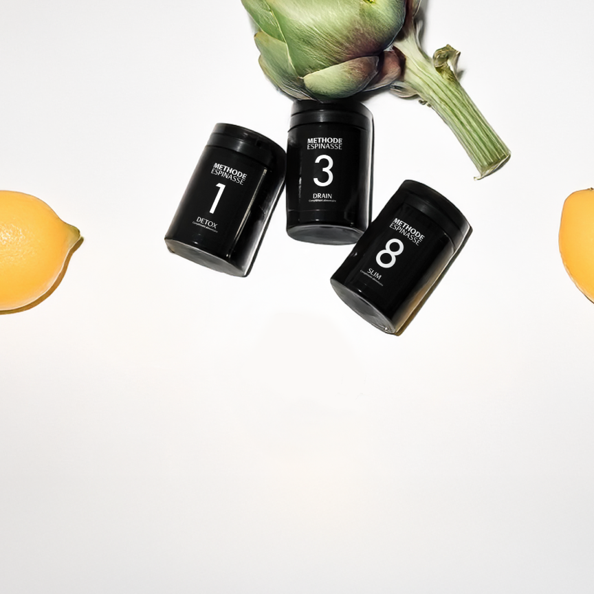
Découvrez 08 - Slim
Your ally for a slimmer figure and healthy weight

Journal

What if the next frontier in skin rejuvenation is not simply about delivering active ingredients to the skin — but influencing the messages skin cells send to one another?
Over the past few years, one word has rapidly moved from regenerative medicine into aesthetic treatments and anti-aging skincare:
exosomes.
Exosome serums.
Exosome skincare.
Exosome facials.
Exosomes after laser treatments.
Exosomes with microneedling.
Stem cell-derived exosomes.
Plant exosomes.
Exosome therapy.
The promises are substantial: stimulating collagen, supporting skin regeneration, reducing wrinkles, repairing UV-induced damage, decreasing inflammation, improving hydration and even “rejuvenating” communication between aging cells.
But behind the marketing fascination lies a biological reality that is far more interesting.
Our cells do not exist in isolation.
They communicate.
Constantly.
And part of that communication can occur through microscopic membrane-bound structures known as:
extracellular vesicles — or EVs.
These vesicles can transport biological material from cells into their surrounding environment and participate in cell-to-cell communication [1].
This ability has made EVs an increasingly important area of research across cell biology, regenerative medicine, wound healing and, more recently, skin aging and skin rejuvenation.
But before asking whether exosomes can regenerate aging skin, we need to clarify something fundamental.
Because much of what is marketed today as:
“exosomes”
may not necessarily meet the strict scientific definition of an exosome.
To understand exosomes, we first need to understand a broader biological category:
extracellular vesicles.
According to the MISEV2023 guidelines from the International Society for Extracellular Vesicles — ISEV — an extracellular vesicle is a particle naturally released from a cell, enclosed by a lipid bilayer and unable to replicate on its own [1].
In simplified terms, an EV is a:
membrane-bound biological particle.
But that does not mean every extracellular vesicle is an exosome.
The term exosome historically refers to a subtype of extracellular vesicle associated with a specific intracellular biogenesis pathway involving the endosomal system and multivesicular bodies.
And that distinction matters.
Not necessarily.
MISEV2023 specifically recommends caution when using biogenesis-based terms such as:
exosome
and
ectosome.
Why?
Because proving the precise subcellular origin of every isolated vesicle population can be extremely difficult [1].
A preparation may contain particles within a size range commonly associated with exosomes.
It may express markers frequently associated with extracellular vesicles.
It may have been isolated from conditioned media.
But none of those characteristics alone necessarily proves that every particle in the preparation originated through the endosomal pathway required to classify it strictly as an exosome.
For that reason, ISEV generally recommends using the broader term:
extracellular vesicles — EVs
when precise biogenesis has not been demonstrated [1].
This may sound like an extremely technical distinction.
It is not.
Because “exosome” has become a marketing term.
In biology, however, it should first describe:
an experimentally supported biological identity.
Throughout this article, we will therefore use EV whenever the available evidence does not justify identifying a preparation specifically as exosomes.
When discussing a study whose authors explicitly use the term “exosomes,” we will preserve their terminology while keeping this distinction in mind.
Because cells are constantly communicating with their environment.
For decades, cell communication was largely understood through soluble signaling molecules such as:
hormones,
cytokines,
growth factors,
neurotransmitters.
A cell releases a signal.
The signal travels.
It encounters another cell.
It may bind to a receptor.
And it changes that cell’s behavior.
Extracellular vesicles have added another layer to our understanding of biological communication.
Instead of releasing only an individual signaling molecule, a cell can release a membrane-bound structure capable of transporting multiple biological components.
One way to visualize it is as:
a biological package sent from one cell to another.
The analogy is imperfect.
But it helps explain why EVs have generated so much scientific interest.
Their composition can vary dramatically depending on the cell that produced them, the biological conditions surrounding that cell and the methods used to isolate the vesicles.
EVs may contain:
proteins;
lipids;
RNA;
and other biologically active molecules [1].
Some EV populations can carry messenger RNA and different types of small regulatory RNA, including microRNAs.
In other words, an EV is not simply an empty bubble.
It can function as a carrier of biological information.
When a recipient cell interacts with an extracellular vesicle or its cargo, that interaction can potentially influence:
gene expression,
inflammation,
oxidative stress,
cell proliferation,
cell migration,
extracellular matrix production.
The outcome depends entirely on the biological context.
And this is precisely why talking about “exosomes” as if they were one standardized skincare ingredient is scientifically problematic.
This distinction is fundamental.
When we talk about:
retinol,
vitamin C,
niacinamide
or even a defined peptide such as:
Acetyl Hexapeptide-8,
we are talking about molecules whose chemical identities can be precisely characterized.
An extracellular vesicle preparation is fundamentally different.
Its biological profile can depend on:
the cell type that produced it;
the physiological state of that cell;
culture conditions;
the culture medium;
collection methods;
isolation methods;
purification;
storage;
characterization of the final preparation.
MISEV guidelines emphasize the importance of precisely these pre-analytical and experimental variables [1].
Two products both marketed as:
“exosome serums”
could therefore be biologically very different.
They may come from different sources.
Contain different vesicle populations.
Have different levels of purity.
Carry different biological cargo.
And use completely different formulations.
So saying:
“exosomes do X”
is often too simplistic.
The more scientifically meaningful questions are:
Which vesicles? Produced by which cells? Under what conditions? Isolated and purified how? Carrying what? Delivered by which route? And at what dose?
Because a large part of regenerative medicine research has focused on mesenchymal stem/stromal cells — MSCs and their effects on tissue repair.
Historically, some of these effects were thought to depend primarily on the cells themselves:
their ability to engraft,
differentiate,
or replace damaged cells.
That understanding has evolved.
A significant part of their biological activity may instead be mediated through paracrine signaling — the signals these cells release into their surrounding environment.
Those signals include soluble molecules.
But they also include extracellular vesicles.
This has led to an intriguing idea:
What if some of the biological effects associated with cells could be harnessed without administering the cells themselves — by using some of the messages they release?
This is one reason mesenchymal stem cell-derived extracellular vesicles have become such an active area of research.
Because skin aging is not simply a structural problem.
It is also a problem of cellular communication.
Young-looking, healthy skin is not simply skin with:
“more collagen.”
It depends on a dynamic network involving:
keratinocytes,
fibroblasts,
immune cells,
melanocytes,
endothelial cells,
the extracellular matrix,
inflammatory mediators,
growth factors,
oxidative stress,
mechanical signaling.
As skin ages and accumulates environmental exposure, those communication networks change.
Some cells become senescent.
Oxidative stress increases.
Inflammatory signaling can shift.
Repair mechanisms become less efficient.
The extracellular matrix is progressively remodeled.
And the balance between:
collagen synthesis
and
collagen breakdown
can change.
This is where extracellular vesicles become particularly interesting.
Because instead of targeting a single molecule, they could theoretically influence multiple signaling networks at once.
Within the dermis, one of the key cell populations is:
fibroblasts.
Fibroblasts help produce and remodel the extracellular matrix.
That matrix includes:
collagens;
elastin;
glycosaminoglycans;
and numerous structural and regulatory proteins.
With intrinsic aging and photoaging, fibroblast biology changes.
Their environment changes.
The collagen network becomes increasingly fragmented.
Mechanical signals change.
And some fibroblasts can enter a state known as:
cellular senescence.
A senescent cell is a cell that has entered a state of stable cell-cycle arrest in response to different forms of stress while remaining metabolically active.
That distinction matters.
Senescent does not mean dead.
The cell remains present.
And it can continue communicating with its environment.
Some senescent cells develop what is known as the:
senescence-associated secretory phenotype — SASP.
They can release mediators capable of influencing:
inflammation,
the extracellular matrix,
neighboring cells,
and the surrounding tissue microenvironment.
Cellular senescence is complex and can also serve useful physiological functions in certain contexts.
But the accumulation of senescent cells with age is considered one of the biological processes involved in tissue aging.
For skin, that raises a fascinating question:
Can we change the signals received by an aging fibroblast?
Several preclinical studies suggest that specific stem cell-derived EV preparations can influence biological parameters associated with fibroblast aging.
In one study involving human dermal fibroblasts, adipose-derived stem cell exosomes were associated with increased type I collagen expression and reductions in several markers associated with oxidative stress and cellular senescence [2].
Researchers observed changes involving:
reactive oxygen species — ROS;
SA-β-gal;
p53;
p21;
p16 [2].
These findings are interesting because they suggest that the experimental effect may extend beyond simply:
“making more collagen.”
The biological state of the fibroblast itself may also be affected.
But there is an essential limitation:
a fibroblast in a laboratory dish is not human skin.
An in vitro experiment demonstrates biological plausibility.
It does not establish clinical anti-aging efficacy.
To understand why extracellular vesicles may be relevant to skin rejuvenation, we need to understand one of the central mechanisms of photoaging.
When skin is exposed to ultraviolet radiation, multiple biological processes are activated.
One of them is the production of:
reactive oxygen species — ROS.
ROS are not inherently harmful.
They also participate in normal physiological signaling.
The problem arises when oxidative balance shifts toward levels capable of damaging cellular components and disrupting signaling pathways.
In the context of skin photoaging, UV exposure can contribute to:
oxidative stress;
inflammation;
DNA damage;
altered cellular signaling;
increased activity of certain matrix metalloproteinases — MMPs.
And MMPs are particularly important when we talk about collagen and wrinkles.
Matrix metalloproteinases, or MMPs, are a family of enzymes involved in the remodeling and degradation of extracellular matrix components.
They perform essential physiological functions.
But increased or dysregulated MMP activity in certain contexts can contribute to excessive matrix degradation.
In photoaged skin, specific MMPs are involved in collagen remodeling and breakdown.
One part of this process can therefore be simplified as:
UV exposure → ROS and cellular signaling → increased activity of certain MMPs → increased extracellular matrix degradation → disruption of the collagen network.
The biology is considerably more complex than this simplified pathway.
But it explains why studies investigating skin photoaging frequently measure:
ROS;
MMP-1;
MMP-3;
type I collagen.
Some experimental evidence suggests they may.
In a model of human fibroblasts exposed to UVB radiation, bone marrow mesenchymal stem cell-derived exosomes were associated with reductions in several markers of oxidative stress and inflammation [3].
Researchers also observed:
decreased MMP-1;
decreased MMP-3;
and
increased type I collagen expression [3].
That profile is particularly interesting in the context of photoaging.
Because it suggests potential activity on both sides of the equation:
fewer signals associated with matrix degradation
and
more signals associated with extracellular matrix maintenance.
But again, the critical word is:
potential.
These experiments do not mean that a skincare serum with the word “exosomes” on its label will automatically reproduce those effects in human facial skin.
This is now one of the most common claims surrounding exosome skincare.
And the scientific answer exists on several levels.
LEVEL 1 — BIOLOGICAL PLAUSIBILITY
Yes.
Cell-based studies show that certain EV preparations can alter the expression of genes and proteins associated with the extracellular matrix, including collagen [2,3].
LEVEL 2 — TISSUE AND ANIMAL MODELS
Research in regenerative medicine provides additional evidence suggesting that EVs can influence tissue repair and remodeling.
LEVEL 3 — HUMAN SKIN
This is where the evidence becomes more limited and heterogeneous.
And this third level should determine how strongly we communicate an aesthetic or skincare claim.
Saying:
“Certain EV preparations increased type I collagen expression in cultured human fibroblasts”
is scientifically defensible when supported by the relevant experiment.
Saying:
“Exosomes rebuild the collagen in your skin”
is a much larger extrapolation.
Because an extracellular vesicle does not necessarily carry a single signal.
This is one of the major conceptual differences between EVs and a single molecular active.
A retinoid can have complex biological effects, but it remains a defined molecule.
A peptide such as Argireline has a defined amino acid sequence.
An EV preparation may contain a heterogeneous population of vesicles carrying multiple biological components.
That creates the possibility of:
multi-target activity.
But it also creates a major scientific challenge.
The more complex the cargo, the more difficult it becomes to determine:
which molecule is responsible for which effect.
And the more difficult it becomes to standardize one preparation against another.
Certain extracellular vesicles can carry or be associated with proteins involved in cellular signaling.
But reducing an EV to:
“a capsule filled with growth factors”
would be inaccurate.
The two concepts need to remain distinct.
Growth factors are signaling molecules capable of influencing cellular behavior.
Depending on the factor and biological context, they may participate in:
cell proliferation,
migration,
differentiation,
angiogenesis,
tissue repair,
extracellular matrix production.
Extracellular vesicles, by contrast, are membrane-bound structures capable of transporting different forms of cargo.
An EV can therefore participate in cellular signaling without itself being a growth factor.
Different growth factor families appear throughout dermatology and regenerative medicine research, including:
EGF — Epidermal Growth Factor;
FGF — Fibroblast Growth Factors;
PDGF — Platelet-Derived Growth Factor;
VEGF — Vascular Endothelial Growth Factor;
TGF-β — Transforming Growth Factor Beta.
But their functions cannot be reduced to:
“growth factor = regeneration.”
Each pathway has its own:
receptors,
target cells,
biological effects,
regulatory mechanisms.
And the same growth factor may produce very different effects depending on:
concentration,
tissue,
timing,
physiological state,
and the presence of other signals.
In biology:
more signaling does not automatically mean better signaling.
Skincare marketing frequently blurs these four concepts.
Scientifically, they are not interchangeable.
EXTRACELLULAR VESICLES / EVs
Membrane-bound particles released by cells that can carry different biological components [1].
EXOSOMES
A subtype of extracellular vesicle associated with a specific biogenesis pathway. The term should ideally be reserved for situations in which that origin has been demonstrated [1].
GROWTH FACTORS
Signaling molecules capable of modulating different cellular functions.
SECRETOME
The much broader collection of substances secreted by a cell into its environment.
CONDITIONED MEDIA
Culture media in which cells have been grown and which may therefore contain a variety of products resulting from cellular secretion.
Conditioned media can contain:
soluble proteins,
cytokines,
growth factors,
metabolites,
extracellular vesicles,
and other components.
A product derived from conditioned media is therefore not automatically:
“an exosome product.”
And an extracellular vesicle preparation is not automatically:
“a growth factor cocktail.”
Because a product can borrow the scientific language of stem cells, exosomes and regenerative medicine without necessarily resembling the preparation used in a published scientific study.
A study may investigate:
isolated and characterized extracellular vesicles derived from human adipose stem cells.
A commercial product may contain:
conditioned media filtrate.
Another may contain:
plant-derived vesicles.
Another:
synthetic liposomes carrying active ingredients.
And another may use the phrase:
“exosome technology”
without giving consumers enough information to determine exactly what the formula contains.
These products cannot be considered scientifically interchangeable.
We have now reached the critical question.
The laboratory data are intriguing.
But we encountered exactly the same issue when examining peptides:
A molecule or biological structure can produce a remarkable effect when placed directly in contact with a cell.
Human skin introduces an additional challenge:
the target has to be reached.
And that challenge may be even greater for a complex extracellular vesicle than for many small cosmetic molecules.
The outermost layer of the epidermis:
the stratum corneum
is specifically designed to restrict the entry of substances from the outside environment.
So an unavoidable question emerges:
How can a complex nanoscale vesicle applied to the surface of the skin reach cells located deeper within the tissue?
This is where the human clinical literature becomes particularly revealing.
Because many of the most interesting studies have not simply applied EVs to intact skin.
Researchers have used:
microneedling.
A prospective, randomized, split-face study published in 2023 evaluated a solution containing human adipose tissue stem cell-derived exosomes combined with microneedling in:
28 participants with signs of facial skin aging [4].
The split-face design is particularly useful.
Each participant partially serves as their own control.
On one side of the face:
microneedling + exosome-containing solution.
On the other:
microneedling + normal saline.
Participants underwent:
three treatment sessions
at:
three-week intervals
and were followed for:
12 weeks [4].
The researchers did not simply ask participants:
“Does your skin look better?”
They used several instruments to objectively assess:
wrinkles;
elasticity;
hydration;
pigmentation [4].
Histopathological analyses were also performed.
At the end of follow-up, the side treated with the exosome-containing solution plus microneedling showed greater improvements across several parameters than the control side [4].
Instrumental measurements showed better outcomes involving:
wrinkles;
elasticity;
hydration;
pigmentation [4].
Overall aesthetic improvement scores were also significantly better on the treated side.
Histopathological findings were consistent with the clinical observations [4].
No serious adverse events were reported during the study [4].
That is genuinely interesting human evidence.
But it immediately raises the right scientific question.
No.
The study demonstrates something much more specific:
In this protocol, an exosome-containing solution derived from human adipose tissue stem cells used in combination with microneedling produced better outcomes than microneedling plus normal saline in 28 participants [4].
That is not the same claim as:
“Applying exosomes to intact skin reduces wrinkles.”
Why?
Because microneedling changes the skin barrier.
It creates microchannels.
It triggers its own biological repair response.
And it can dramatically change the way a topical preparation interacts with underlying tissue.
There are therefore two variables:
the biological preparation
and
the delivery technology.
And that distinction may prove central to the future of extracellular vesicles in aesthetic medicine.
Possibly.
This is one of the most interesting hypotheses emerging from the current literature.
Extracellular vesicles may have fascinating biology.
They may carry complex biological cargo.
They can modify cellular behavior in experimental models.
But a technology only becomes clinically meaningful if the biological signal can:
reach the right cell,
in the right place,
in a functional state,
at a biologically relevant dose.
This is why EV preparations are increasingly being studied alongside technologies such as:
microneedling;
radiofrequency microneedling;
fractional laser;
transdermal delivery systems.
And this brings us back to the exact same principle we encountered when investigating Argireline:
the mechanism is only the first half of the story.
The second half is:
delivery.
Next, we move further away from the Petri dish and deeper into the human clinical evidence.
Exosomes + microneedling.
Exosomes + radiofrequency.
Exosomes + fractional laser.
Wrinkles.
Elasticity.
Hydration.
Pigmentation.
Acne scars.
Skin repair.
We will examine why some human studies are genuinely promising — and why their methodology still does not establish extracellular vesicles as a definitively proven anti-aging technology.
We will also investigate one of the most controversial areas of exosome skincare:
“plant exosomes” and plant-derived extracellular vesicles.
Do plants really produce the same “exosomes” as human cells?
Then:
Do topical growth factors actually work?
What is the difference between an exosome product, conditioned media and a growth factor serum?
Can these biological structures actually penetrate the skin?
And, crucially:
What do we know about exosome safety?
Because once we move from:
application to intact skin
to:
microneedling, laser-assisted delivery or deeper administration,
the question is no longer simply about efficacy.
It becomes a question of:
purity, characterization, sterility, standardization and regulation.
Finally, we will compare the current enthusiasm with the most recent systematic reviews to answer the central question:
Are exosomes really the next frontier in skin regeneration — or is science still catching up with a market that has already embraced the word?
This is where the most important part of the investigation begins.
In laboratory models, certain extracellular vesicles can influence fibroblast behavior, oxidative stress, MMP activity, cellular senescence and collagen expression.
But one fundamental question remains:
Do those effects actually translate to human skin?
Clinical evidence is beginning to accumulate.
And it is compelling enough to explain the growing scientific interest in exosomes for skin rejuvenation.
But there is one critical detail.
Many of the most encouraging results have not been obtained by simply applying:
an exosome serum to intact skin.
Instead, extracellular vesicles have frequently been combined with technologies designed to modify or partially bypass the skin barrier:
That distinction significantly changes how we should interpret the evidence.
As discussed in Part 1, a prospective, randomized, split-face study evaluated 28 participants with signs of facial skin aging [4].
After three treatment sessions spaced three weeks apart, the side treated with microneedling plus a solution containing human adipose tissue stem cell-derived exosomes showed better results than the side treated with microneedling plus normal saline.
Improvements included:
wrinkles;
elasticity;
hydration;
pigmentation [4].
Importantly, the outcomes were not based solely on subjective assessments.
Instrument-based measurements were used, along with histological analyses.
But the study also had limitations.
It included only 28 participants.
The authors noted that because previous data were insufficient to perform a formal power calculation, sample size was determined based on feasibility [4].
And most importantly:
the study tested a combination treatment.
It does not tell us what the same exosome preparation would have achieved when simply applied to intact skin without microneedling.
A randomized split-face study published in 2026 provides another particularly interesting piece of evidence [5].
The study included:
16 adults age 40 and older
with visible signs of facial aging.
On one side of the face, researchers used a solution containing human adipose tissue-derived stem cell exosomes combined with:
ultrasound-assisted transdermal delivery.
The other side received normal saline using the same delivery protocol [5].
Three treatments were performed at two-week intervals.
Researchers assessed:
transepidermal water loss — TEWL;
stratum corneum hydration;
multiple elasticity parameters;
wrinkles;
pigmentation;
as well as exploratory parameters involving the skin microbiome [5].
The results suggested improvements in skin barrier function, including decreased TEWL and favorable changes in certain hydration and elasticity measurements [5].
This is interesting.
But again:
16 participants.
And again:
assisted transdermal delivery.
A pattern is beginning to emerge.
Possibly.
Because many of the most interesting human studies are not simply testing:
EVs vs. nothing.
They are testing:
EVs + a delivery procedure
against:
the procedure alone or another control.
That is a major methodological distinction.
The stratum corneum is a remarkably effective barrier.
And unlike many small cosmetic molecules, extracellular vesicles are complex biological structures.
Their size may be measured on the nanoscale, but:
nanoscale does not automatically mean transdermal.
Size alone does not determine skin penetration.
Membrane composition, surface charge, stability, interactions with skin lipids, formulation and barrier integrity may all matter.
So the question is not simply:
“Do exosomes work?”
It is:
“How can functional extracellular vesicles be delivered to the cells we actually want to influence?”
Using extracellular vesicles after procedures that create a controlled disruption of the skin barrier is another major area of research.
A prospective, randomized, double-blind, split-face trial included 25 patients with atrophic acne scars [6].
All participants underwent three sessions of:
fractional CO2 laser treatment.
After each session, one side of the face received a gel containing human adipose tissue stem cell-derived exosomes.
The other side received a control gel.
After 12 weeks, the clinical scar score had decreased by:
32.5% on the exosome-treated side
versus:
19.9% on the control side [6].
Researchers also reported:
less erythema
and
shorter downtime
on the exosome-treated side [6].
The findings are compelling.
But it is critical to understand what this study actually demonstrates.
This trial does not demonstrate that:
“Exosomes reduce wrinkles by 32.5%.”
The participants had:
atrophic acne scars.
And the clinical endpoint involved scar improvement.
The study is nevertheless relevant when considering the potential role of extracellular vesicles in:
repair;
tissue remodeling;
post-procedure recovery.
It also raises an important hypothesis:
EVs may be particularly useful when applied in a skin environment temporarily made more accessible by an aesthetic procedure.
That is very different from conventional anti-aging skincare.
Because these procedures trigger biological responses of their own.
Microneedling and fractional lasers can create controlled tissue injury.
That injury initiates a repair cascade involving:
inflammation,
cellular mediators,
cell migration,
fibroblasts,
extracellular matrix remodeling.
At the same time, temporarily altering the skin barrier may allow certain substances to interact with deeper layers more readily.
The strategy may therefore involve intervening during a:
biological window of repair.
But this also makes the results more difficult to interpret.
When:
laser + EVs
performs better than:
laser alone,
it is reasonable to infer an additional contribution from the preparation being tested.
But we cannot automatically extrapolate that difference to:
EVs alone on intact skin.
This is precisely where the current evidence is less definitive.
Topical formulations are beginning to be studied.
But the available trials tend to be:
small;
short;
heterogeneous;
and they do not all investigate the same types of extracellular vesicles.
Formulation is another major challenge.
An EV placed inside a skincare product needs to retain characteristics compatible with the intended biological activity despite:
manufacturing,
purification,
formulation,
storage,
transport,
temperature,
shelf life.
An impressive study involving freshly isolated EVs in a research laboratory is therefore not automatically evidence that a commercial exosome face serum will produce the same biological effect.
A 2026 systematic review specifically evaluated extracellular vesicle-based therapies for nonsurgical facial rejuvenation [7].
It included:
16 studies
representing:
503 participants [7].
The studies used a variety of designs, including:
split-face trials,
comparative studies,
single-arm studies.
Most reported improvement in at least one outcome, including:
wrinkles;
pigmentation;
skin hydration;
skin elasticity;
overall appearance [7].
In comparative studies, EV-treated areas generally showed favorable outcomes.
No serious adverse events were reported in the included studies [7].
At first glance, the evidence looks highly encouraging.
But the second half of the analysis matters even more.
The systematic review assessed both risk of bias and certainty of evidence [7].
For most efficacy outcomes, risk of bias was considered:
moderate to critical.
Using the GRADE framework, overall certainty of evidence was rated:
low or very low [7].
Why?
Several recurring limitations remain:
small sample sizes,
heterogeneous protocols,
different EV sources,
different preparation methods,
different delivery technologies,
different outcome measures,
relatively short follow-up periods,
frequent lack of blinding or robust controls.
This may be the most accurate summary of the science today:
The clinical signals are promising.
But:
the definitive evidence is not there yet.
The word:
“rejuvenation”
is extremely broad.
Current human studies allow us to discuss improvements in specific parameters within specific protocols:
wrinkles;
hydration;
elasticity;
pigmentation;
skin barrier function;
recovery after certain procedures.
They do not establish that extracellular vesicles:
biologically reverse human aging.
And they certainly do not demonstrate that an exosome facial or serum:
“makes skin cells years younger.”
Claims like these go far beyond the evidence currently available.
It is an appealing phrase.
But it should be used with extreme caution.
Extracellular vesicles can carry biological signals capable of altering gene expression and influencing the behavior of recipient cells.
That is precisely what makes them so fascinating.
But:
changing a signaling pathway is not the same as cellular reprogramming in the strict biological sense.
Cellular reprogramming has a much more specific meaning in biology.
Describing an exosome skincare treatment as:
“cellular reprogramming”
would therefore be an overstatement based on current evidence.
This is one of the fastest-growing — and most confusing — areas of next-generation skincare.
Plants also produce vesicular structures.
The scientific literature frequently describes them as:
plant-derived extracellular vesicles
or:
extracellular vesicle-like particles.
Automatically applying the term:
“exosome”
to every vesicular nanoparticle isolated from a plant creates the same nomenclature problem we encounter with human-derived EVs.
Classification should depend on appropriate characterization and, ideally, demonstrated biogenesis.
For this reason:
“plant-derived extracellular vesicles”
is often more scientifically precise than:
“plant exosomes.”
Recent human studies have begun investigating skincare formulations containing Centella asiatica-derived extracellular vesicles.
A 2025 study evaluated an ampoule containing Centella-derived EVs in 20 female participants over two weeks [8].
Researchers reported changes across several instrumentally measured parameters, including:
pores,
skin roughness,
wrinkle depth,
hydration,
skin density [8].
Another 28-day pilot study evaluating a Centella EV-based skincare formulation also reported improvements in hydration, elasticity and several measures of skin texture [9].
These findings are interesting.
But we need to examine the entire formulation.
In one of these studies, the tested formulation did not contain plant-derived EVs alone [8].
It combined the EV preparation with other cosmetic ingredients.
Therefore, if the product improves a parameter, we can conclude:
“The formulation produced an improvement.”
We cannot automatically conclude:
“The extracellular vesicles alone caused the improvement.”
This is a fundamental principle in skincare research.
A clinical trial on a formula is not necessarily a clinical trial on an individual ingredient.
That distinction will become increasingly important as the number of plant exosome skincare products continues to grow.
Not directly.
The fact that two structures fall within the broad category of extracellular vesicles does not mean they carry the same biological information.
Their cargo depends on the organism and cell of origin.
An EV derived from:
a human mesenchymal stem/stromal cell
is not biologically equivalent to an EV derived from:
Centella asiatica.
The shared term:
EV
describes a category of structures.
It does not guarantee identical cargo or biological activity.
Just as the word:
“peptide”
does not mean every peptide produces the same effect.
Scientifically, this comparison does not make much sense.
A peptide is generally a molecule composed of a sequence of amino acids.
An extracellular vesicle is a complex membrane-bound structure capable of carrying multiple biological components.
The two technologies do not necessarily act:
on the same targets,
through the same pharmacology,
at the same depth,
or according to the same biological mechanisms.
There is therefore no scientifically valid hierarchy such as:
retinol < peptides < exosomes.
A technology that sounds more sophisticated is not automatically more effective.
Growth factors have been studied in skincare for considerably longer than exosomes.
They can participate in mechanisms involving:
cell proliferation,
migration,
differentiation,
wound healing,
extracellular matrix biology.
Topical preparations containing different growth factors have been investigated for facial skin aging and rejuvenation.
And their clinical literature is currently broader than that of extracellular vesicles.
A 2023 systematic review evaluated the evidence for topical growth factor preparations in facial skin rejuvenation [10].
It identified:
33 studies
including:
9 randomized controlled trials
and:
24 uncontrolled case series.
In total:
1,180 participants
were treated with:
23 different growth factor-containing preparations [10].
That is a substantially larger body of evidence than currently exists for extracellular vesicles.
But more evidence does not automatically mean better evidence.
Overall, the studies reported improvements in various parameters associated with photoaging and skin appearance [10].
But the systematic review also identified considerable heterogeneity.
The formulations differed.
The sources of the growth factors differed.
Protocols differed.
Treatment durations differed.
And most of the studies were small: 28 of the 33 studies included 40 participants or fewer [10].
The evidence therefore supports growth factors as an interesting field in anti-aging skincare research.
It does not establish every growth factor serum as equivalent.
Because growth factors are generally relatively large proteins.
And human skin is not designed to allow large biologically active proteins to freely cross its outer barrier.
We therefore encounter the same fundamental problem:
Having a biologically powerful signal is not enough.
You still have to:
stabilize it;
deliver it;
get it to the right biological target.
This is why advanced formulation technologies and procedures that temporarily alter the skin barrier also appear throughout the growth factor literature.
A simplified distinction is:
GROWTH FACTOR
→ a molecular signal.
EXTRACELLULAR VESICLE
→ a biological transport system capable of carrying multiple types of signals.
An EV may theoretically transport:
proteins,
lipids,
RNA,
and different molecular regulators.
It is this potential for complex biological communication that makes extracellular vesicles so compelling.
But complexity cuts both ways.
It potentially increases:
biological richness
while also increasing:
the challenge of standardization.
Imagine two batches of a conventional skincare active.
If the active is a chemically defined molecule, it is possible to precisely verify:
identity,
purity,
concentration.
With an extracellular vesicle preparation, the situation is much more complex.
Researchers and manufacturers may need to characterize:
the biological source,
culture conditions,
particle size,
particle concentration,
markers,
contaminants,
associated proteins,
nucleic acids,
isolation methods,
stability.
Changing the conditions in which the producer cells are grown may potentially change what those cells secrete.
In other words, the cell itself becomes:
part of the manufacturing process.
That is fundamentally different from a conventional chemically defined skincare ingredient.
We do not currently have enough evidence to say so.
Establishing a meaningful dose-response relationship would require studies comparing multiple clearly characterized doses under otherwise comparable conditions:
same source,
same preparation,
same formulation,
same delivery route,
same population,
same outcome measures.
Those data remain limited.
So the most impressive number on an exosome serum:
“1 billion exosomes”
or
“10 billion particles”
does not, by itself, predict clinical efficacy.
Counting particles is not the same as measuring biological activity.
No.
Two preparations containing the same number of particles could theoretically differ dramatically in:
source,
purity,
cargo,
biological activity,
stability.
There is also a distinction between:
total particle count
and
the number of biologically functional vesicles.
A huge number on a skincare label may sound impressive.
It does not replace:
biological characterization.
There is no universal answer.
The question should always be:
Which extracellular vesicles, from what source, prepared how and administered by which route?
In the small aesthetic studies currently available, no major short-term safety signal has emerged across the protocols included in the recent systematic review [7].
But that does not establish that:
“exosomes are universally safe.”
The populations studied remain limited.
Follow-up periods are generally short.
And the products themselves are highly heterogeneous.
This is probably the single most important safety distinction.
Applying a preparation:
to intact skin
is not biologically equivalent to applying it:
immediately after microneedling or laser.
And neither is equivalent to:
injecting it.
Once the skin barrier is bypassed, additional issues become critical:
sterility;
endotoxins;
microbial contamination;
purity;
immunogenicity;
composition;
dose;
biodistribution.
A cosmetic product intended for topical use does not automatically become suitable for intradermal administration simply because it contains extracellular vesicles.
In the United States, the FDA currently states that there are:
no FDA-approved exosome products [11].
The agency explains that exosome products intended to treat diseases or conditions in humans fall within its regulatory oversight and generally require appropriate FDA authorization [11].
The FDA has also issued public safety notifications following reports of serious adverse events involving certain unapproved products marketed as containing exosomes [12].
This needs to be interpreted correctly.
It does not mean:
“All exosome skincare is dangerous.”
It means:
“A biologically interesting technology is not automatically an approved medical therapy.”
And it reinforces why route of administration matters.
No.
A cosmetic formulated for topical application should never be assumed to be suitable for injection.
Composition, manufacturing requirements and sterility standards are not the same.
The same caution applies when products are used after procedures that create microchannels in the skin.
The words:
“exosome serum”
do not guarantee that a product was designed, manufactured or clinically evaluated for delivery beyond the intact skin barrier.
This question is much more complex than a simple yes or no.
Extracellular vesicles naturally participate in cell-to-cell communication.
In different biological contexts, EVs can carry signals involved in:
proliferation,
angiogenesis,
immunity,
inflammation.
Cancer cells themselves also produce extracellular vesicles and use them to communicate within the tumor microenvironment.
That does not mean a cosmetic EV preparation:
“causes cancer.”
The available evidence does not support such a blanket conclusion.
But it does explain why any technology designed to deliver biologically active signals needs to be:
characterized;
standardized;
toxicologically evaluated;
and studied according to its intended route of administration.
The term:
“cell-free”
does not mean:
“biologically inert.”
Based on the evidence available today:
Current evidence does not establish that:
Calling exosomes a:
“revolution”
is probably premature.
But dismissing extracellular vesicles as nothing more than a skincare trend would be equally reductive.
The biology behind them is real.
And it is fascinating.
For decades, skincare has primarily focused on delivering:
molecules.
Antioxidants.
Retinoids.
Acids.
Vitamins.
Peptides.
Extracellular vesicles potentially represent a different paradigm.
Instead of delivering only:
a molecule,
the goal becomes harnessing:
a natural system of cell-to-cell communication.
That is a major conceptual shift.
Possibly.
One of the fundamental principles of modern biology is that a cell is not defined only by the nutrients it receives.
It is also shaped by:
the signals it receives.
Hormones.
Cytokines.
Growth factors.
Neurotransmitters.
Metabolites.
Extracellular vesicles.
Mechanical signals.
Aging, therefore, is not simply a loss of structural material.
It also involves progressive changes in:
biological information
and in the way cells respond to that information.
This is precisely what makes extracellular vesicles so compelling.
Exosomes may be one of the clearest examples of how quickly a discovery from fundamental cell biology can become a global aesthetic trend.
First:
a mechanism of intercellular communication.
Then:
a potential regenerative medicine platform.
And now:
a term printed on skincare serums, microneedling protocols and aesthetic treatments.
But there is a considerable scientific distance between those three worlds.
Extracellular vesicles are real.
Their ability to carry biological information is real [1].
Their capacity to influence cellular behavior in experimental models is real.
And some human studies have reported promising improvements involving:
wrinkles;
skin elasticity;
hydration;
pigmentation;
recovery after certain aesthetic procedures [4–7].
But the current science requires one essential qualification:
We cannot yet turn those findings into a universal claim about “exosomes.”
Because there is no single:
“exosome.”
There are populations of extracellular vesicles.
Produced by different cells.
Under different conditions.
Isolated using different methods.
Purified differently.
Carrying different cargo.
Formulated differently.
And delivered through different routes.
That is why the question:
“Do exosomes work?”
may ultimately be too simplistic.
The more scientifically meaningful question is:
Which extracellular vesicles, for which indication, at what dose, through which delivery route and with what level of biological characterization?
And that immediately leads to another:
Can a natural system of cellular communication be transformed into a reproducible, stable, standardized and safe skin technology?
If the answer eventually becomes yes, extracellular vesicles could open the door to a new generation of approaches to skin aging and regeneration.
Approaches based not simply on:
the ingredient.
But on:
the biological message.
An exosome is a subtype of extracellular vesicle associated with a specific endosomal biogenesis pathway. When that origin has not been demonstrated, international scientific guidelines generally favor the broader term extracellular vesicle — EV [1].
An extracellular vesicle is a lipid-bilayer-delimited particle naturally released by a cell that cannot replicate on its own [1]. EVs can transport different biological components and participate in cell-to-cell communication.
No.
A stem cell is a living cell.
An extracellular vesicle is a particle released by a cell.
A stem cell-derived EV preparation therefore does not necessarily contain the stem cells themselves.
Depending on their origin and preparation, extracellular vesicles may carry proteins, lipids and different types of nucleic acids [1].
Certain preclinical studies have found that specific EV preparations can increase markers associated with collagen while reducing certain MMPs in fibroblast models [2,3]. This does not mean every commercial exosome serum automatically produces the same effect in human skin.
Some human studies report improvements in wrinkles, particularly when EV-containing preparations are combined with procedures such as microneedling [4]. However, the overall certainty of the clinical evidence remains low to very low [7].
Human evidence for topical formulations is beginning to emerge, including studies involving plant-derived EVs [8,9]. However, trials remain small and formulations can contain multiple active ingredients. More clinical evidence currently exists for protocols combining EVs with delivery technologies.
The ability of different extracellular vesicles to cross intact human skin in biologically meaningful quantities has not been sufficiently characterized to support a broad conclusion. Penetration is likely to depend on the vesicle, formulation and condition of the skin barrier.
A randomized split-face study involving 28 participants found better outcomes with microneedling plus a solution containing adipose tissue stem cell-derived exosomes than with microneedling plus normal saline [4]. Larger trials are needed to confirm the magnitude and reproducibility of the effect.
Some clinical studies have evaluated this strategy. A randomized split-face trial involving fractional CO2 laser for acne scars found greater improvement and faster recovery with an exosome-containing gel than with control treatment [6]. This does not mean every commercial exosome product is appropriate for post-laser use.
Plants produce extracellular vesicles. When their precise biogenesis has not been demonstrated, plant-derived extracellular vesicles is generally a more scientifically cautious term than “plant exosomes.”
Small human studies have reported improvements in certain skin parameters with formulations containing Centella asiatica-derived extracellular vesicles [8,9]. Small sample sizes, short study durations and multi-ingredient formulations limit conclusions about the isolated effects of the EVs themselves.
A growth factor is a signaling molecule. An extracellular vesicle is a membrane-bound structure capable of transporting multiple types of biological molecules and participating in intercellular communication.
The secretome is the broad collection of substances secreted by a cell. Extracellular vesicles may be one component of the secretome, which also includes soluble molecules.
Conditioned media is the culture medium in which cells have been grown. It may contain EVs, soluble proteins, growth factors, cytokines, metabolites and other cell-derived components.
A systematic review of 33 studies involving 1,180 participants found improvements in different facial aging parameters with certain topical growth factor preparations [10]. However, the products and study protocols were highly heterogeneous.
This has not been established. Particle count alone does not reveal the source, purity, cargo, stability or biological activity of an EV preparation.
Available small aesthetic studies are relatively reassuring in the short term, but they cannot establish universal or long-term safety [7]. Biological source, manufacturing, purity and, critically, route of administration all matter.
The FDA currently states that there are no FDA-approved exosome products [11].
A topical cosmetic product should never be assumed to be suitable for injection. In the United States, exosome products intended to treat diseases or conditions are subject to FDA regulatory requirements [11].
After collagen, NAD+, PDRN, signal peptides, Matrixyl®, GHK-Cu, Argireline® and now extracellular vesicles, we can go even deeper into the biology of aging:
CELLULAR SENESCENCE & SENOLYTICS: CAN WE ELIMINATE THE CELLS THAT MAKE US AGE?
Senescent cells.
SASP.
p16.
p21.
Inflammaging.
Fibroblasts.
Mitochondria.
Quercetin.
Fisetin.
Senolytics.
Senomorphics.
And a question increasingly central to longevity science:
Does aging also mean accumulating cells that no longer function as they once did — but refuse to disappear?
A Doctor of Pharmacy and expert in Predictive & Preventive Medicine, Dr. ESPINASSE has spent more than 20 years developing an integrative approach connecting nutrition, metabolism, the microbiome, cellular function, aging and longevity.
Her clinical experience with more than 20,000 patients, supported by more than 15,000 assessments, led her to develop Cellular Nutrition®: an approach that considers nutrients and bioactive compounds through the physiological and cellular mechanisms they are intended to support.
Through METHODE ESPINASSE, Dr. ESPINASSE translates more than two decades of clinical experience into targeted nutritional protocols focused on health, beauty, longevity and healthy aging.
[1] Welsh JA, Goberdhan DCI, O’Driscoll L, et al. Minimal information for studies of extracellular vesicles (MISEV2023): From basic to advanced approaches. Journal of Extracellular Vesicles. 2024;13.
URL: https://pmc.ncbi.nlm.nih.gov/articles/PMC10850029/
[2] Adipose-derived stem cell exosomes and human dermal fibroblast senescence: effects on oxidative stress, senescence-associated markers and collagen expression. PMID: 35813225.
URL: https://pubmed.ncbi.nlm.nih.gov/35813225/
[3] Mesenchymal stem cell-derived exosomes protect human dermal fibroblasts against UVB-induced photoaging: effects on oxidative stress, inflammation, MMP-1, MMP-3 and collagen I. PMID: 36746125.
URL: https://pubmed.ncbi.nlm.nih.gov/36746125/
[4] Park GH, Kwon HH, Seok J, et al. Efficacy of combined treatment with human adipose tissue stem cell-derived exosome-containing solution and microneedling for facial skin aging: A 12-week prospective, randomized, split-face study. Journal of Cosmetic Dermatology. 2023;22(12):3418–3426. DOI: 10.1111/jocd.15872. PMID: 37377400.
URL: https://pubmed.ncbi.nlm.nih.gov/37377400/
[5] Choi BY, Kim HJ, Kim MJ, et al. Adipose tissue-derived stem cell exosomes enhance skin barrier function and show exploratory associations with the skin mycobiome in aging skin. Journal of Microbiology. 2026;64(6). DOI: 10.71150/jm.2603020. PMID: 42403177.
URL: https://pubmed.ncbi.nlm.nih.gov/42403177/
[6] Kwon HH, Yang SH, Lee J, et al. Combination Treatment with Human Adipose Tissue Stem Cell-derived Exosomes and Fractional CO2 Laser for Acne Scars: A 12-week Prospective, Double-blind, Randomized, Split-face Study. Acta Dermato-Venereologica. 2020. DOI: 10.2340/00015555-3666. PMID: 33073298.
URL: https://pubmed.ncbi.nlm.nih.gov/33073298/
[7] Gaebe K, Lowe K, Bosnalie D, et al. Effectiveness and Safety of Extracellular Vesicle-Based Therapies for Nonsurgical Facial Rejuvenation: A Systematic Review. 2026.
URL: https://journals.sagepub.com/doi/10.1177/26893614261457879
[8] Clinical Efficacy and Safety Evaluation of a Centella asiatica (CICA)-Derived Extracellular Vesicle Formulation for Anti-Aging Skincare. Cosmetics. 2025;12(4):135.
URL: https://www.mdpi.com/2079-9284/12/4/135
[9] Chang TM, Wu CC, Huang HC, et al. A 28-Day Pilot Study of the Effects on Facial Skin Hydration, Elasticity, and Texture of a Centella asiatica Extracellular Vesicle-Based Skin Care Formulation. Cosmetics. 2025;12(5):186. DOI: 10.3390/cosmetics12050186.
URL: https://www.mdpi.com/2079-9284/12/5/186
[10] Quinlan DJ, Ghanem AM, Hassan H. Topical growth factor preparations for facial skin rejuvenation: A systematic review. Journal of Cosmetic Dermatology. 2023;22(7):2023–2039. DOI: 10.1111/jocd.15644. PMID: 37222303.
URL: https://pubmed.ncbi.nlm.nih.gov/37222303/
[11] U.S. Food and Drug Administration. Consumer Alert on Regenerative Medicine Products Including Stem Cells and Exosomes.
URL: https://www.fda.gov/vaccines-blood-biologics/consumers-biologics/consumer-alert-regenerative-medicine-products-including-stem-cells-and-exosomes
[12] U.S. Food and Drug Administration. Public Safety Notification on Exosome Products.
URL: https://www.fda.gov/vaccines-blood-biologics/safety-availability-biologics/public-safety-notification-exosome-products